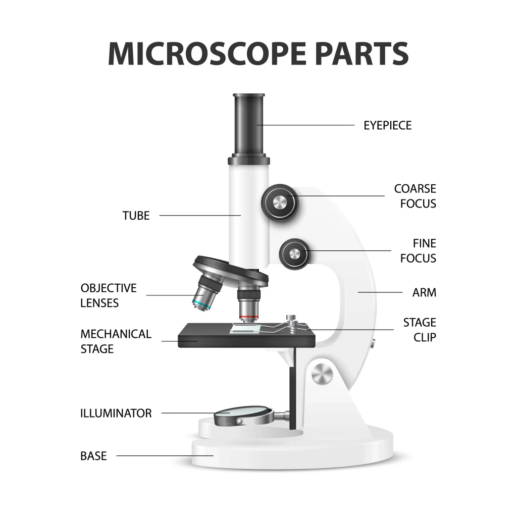
A microscope is an essential tool for scientists, researchers, and medical professionals. It allows us to observe and analyze objects that are too small to be seen with the naked eye. There are several parts of the microscope that are important for its proper functioning.
The main function of a microscope is to provide a magnified view of small objects or organisms, such as bacteria, cells, or tissues. The main parts include the following:

Structural parts of a microscope:
There are three major structural parts of a microscope.
- The head comprises the top portion of the microscope, which contains the most important optical components, and the eyepiece tube.
- The base serves as the microscope’s support and holds the illuminator.
- The arm is the component of the microscope that connects the eyepiece tube to the base of the instrument as well as the head of the microscope. In addition to providing support for the head of the microscope, it is also used for transporting the instrument. Some of the highest-quality microscopes contain an articulated arm that has more than one joint, which enables the tiny head to have greater mobility, resulting in improved vision.

Optical parts of a microscope
There are the following major optical parts of a microscope.
The Eyepiece
The eyepiece, also called the ocular lens, is the lens at the top of a microscope through which the viewer looks. The magnification of the normal eyepiece is 10 times greater. You have the option of exchanging it with an eyepiece that has magnification levels ranging from 5x to 30x.
The Eyepiece tube
The eyepiece lens is placed inside the eyepiece tube. It keeps the eyepiece in a position that’s properly aligned with the objective lenses. It also positions the eyepiece and objective lenses within a certain distance range, resulting in sharp pictures.
Monocular microscopes have one eyepiece. Binocular microscopes include two eyepieces for dual vision. The eyepiece tube may be rotated to match the user’s eye distance. Trinocular microscopes include a third eyepiece tube for a microscope camera.

The objective lenses
These are the lenses that are closest to the object being viewed. They are typically labeled with a number, such as 4x, 10x, or 40x, which indicates the magnification power of the lens.
On a microscope, there are about one to four objective lenses (as shown in the first fig), some of which face backward and others that face forward. Each lens has a unique magnification capability.
The focus knobs
These are the knobs that are used to adjust the focus of the microscope. These Adjustment knobs are of two types: fine adjustment knobs and coarse adjustment knobs.
You should use the coarse focus knob to bring the specimen into approximate or near focus. Then you use the fine focus knob to sharpen the focus quality of the image. When viewing with a high-power objective lens, carefully focus by only using the fine knob.
Stage
This is the part of the microscope where the specimen is placed so it may be examined. The specimen slides are held in place by stage clips that are included with the stage. It is usually adjustable so that the object can be moved and positioned for optimal viewing.
The light source (Illuminator)
This is the source of illumination for the object being viewed. In order to provide a constant level of illumination, halogen bulbs are often used. At this time, LED lights are becoming ever more widespread. Sometimes mirrors are also used instead of a built-in light.
Condenser
Condensers are specialized lenses that are put in place between an illuminator and the specimen in order to gather and concentrate the light. They are located beneath the microscope’s stage adjacent to the diaphragm.
At magnifications of 400x and above, sharp images can only be made with condensers. The image gets sharper as the magnification of the condenser gets higher. For a 40x objective lens, the best condenser is a stage-mounted 0.65 N.A. or greater. For better clarity, if your microscope goes to 1000x or higher, you need a focusable condenser lens with an N.A. of 1.25 or higher.
The condenser focus knob
This is a knob that controls how high or low the condenser is moved, which in turn controls how much light is focused on the specimen.
The diaphragm
The diaphragm is a disc with a series of holes that can be adjusted to control the amount of light that reaches the specimen. By reducing the amount of light, the diaphragm can help to improve the contrast of the image and make it easier to view certain types of specimens.
Functions of different types of microscopes
There are several different types of microscopes, each of which has its own specific functions and uses. For example:
- Compound microscopes are the most common type of microscope and are used for a wide range of applications. They use a combination of lenses to magnify the image and provide high levels of resolution and detail.
- Stereo microscopes, also known as dissecting microscopes, are used for viewing three-dimensional objects, such as insects, plants, or small mechanical parts.
- Confocal microscopes are used for high-resolution imaging of biological samples. They use a laser to scan the sample and produce detailed images with minimal distortion.
- Electron microscopes use a beam of electrons instead of light to produce highly magnified images of very small objects, such as atoms or molecules.
Microscopes are generally used in scientific research, medical diagnosis, and industrial quality control. Scientists and researchers can use them to look at the structure and function of both living and nonliving things on a microscopic scale. This can teach us a lot about how living things work and help us come up with new technologies and treatments.
Parts of a Microscope Questions & Answers (FAQs)
Ans. A microscope is an essential tool for scientists, researchers, and medical professionals. It allows us to observe and analyze objects that are too small to be seen with the naked eye.
Ans. Magnification is the ratio between the height of an image and the height of an item. The overall enlargement of an object’s picture is measured by the microscope’s magnification. The sum of the powers of the eyepiece and objective lenses is the magnification power.
Ans. https://biologynotesweb.com/wp-content/uploads/2022/12/Parts-of-the-microscope-1024×1024.webp
Ans. The coarse adjustment knob is the bigger of the two knobs and is located closest to the arm of the microscope. The fine adjustment knob is the smaller of the two knobs and is located further away from the arm of the microscope.
The coarse adjustment knob moves the stage up and down to bring the specimen into focus. The fine adjustment knob brings the specimen into sharp focus under low power and is used for all focusing when using high-power lenses.
1. Head
2. Arms
3. Base
Ans. The lenses used with a microscope for examination purposes are called “objective” lenses. The objective lens in a stationary microscope then directs the object’s reflected light up a tube and toward the ocular lens, which is the lens the user sees through.
Microscope Parts Worksheet

Key points
I understand. The web page titled “Parts of a Microscope with Labeled Diagram and Functions” has the following key takeaways:
- 🔍 The microscope is an essential tool for scientists, researchers, and medical professionals.
- 🧬 The main function of a microscope is to provide a magnified view of small objects or organisms, such as bacteria, cells, or tissues.
- 🌟 There are several important parts of the microscope that contribute to its proper functioning, including the eyepiece, objective lenses, focus knobs, stage, light source, and condenser.
- 📏 The microscope has three major structural parts: the head, the base, and the arm.
- 📐 Adjustment knobs are used to adjust the focus of the microscope. Coarse adjustment knobs bring the specimen into approximate focus, while fine adjustment knobs sharpen the focus quality of the image.
- 💡 At higher magnifications, condensers are necessary to produce sharp images. The best condenser depends on the magnification of the objective lens, but an N.A. of 1.25 or higher is needed for magnifications of 1000x or higher.
- 🤝 Microscopes are used in scientific research, medical diagnosis, and industrial quality control to study the structure and function of living and nonliving things on a microscopic scale.
References and Sources
- https://rsscience.com/compound-microscope-parts-labeled/
- Microbiology by Lansing M. Prescott (5th Edition)
- https://www.pobschools.org/cms/lib/NY01001456/Centricity/Domain/349/TheMicroscope-howtouse.pdf
- https://sciencing.com/parts-microscope-uses-7431114.html
- https://www.amscope.com/microscope-parts-and-functions/
- https://cpb-us-e1.wpmucdn.com/cobblearning.net/dist/3/4204/files/2018/08/Parts-of-the-Microscope-103b21p.pdf
